Brown Spots on Your Legs: When to Get Them Checked
In women, melanoma forms most often on the arms and legs. The ABCDE signs and the ugly duckling rule show which spots need a dermatologist.
By Rachel Moreno · Published
11 sources · Our standards
Straight answer
Most brown spots on legs are harmless, but some aren’t, and you can’t reliably tell which is which at home. Get a spot checked by a board-certified dermatologist if it shows any of the AAD’s ABCDE warning signs, looks different from your other spots, or is new, changing, itching or bleeding. In women, melanoma forms most often on the arms and legs, and melanoma caught early is highly treatable.
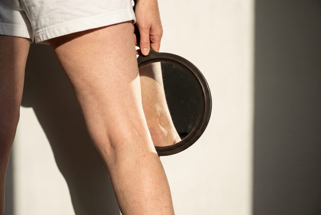
This is a guide to when to book an appointment, not a way to rule anything out yourself. Brown spots on legs can be age spots, scars from old bug bites, harmless growths or, occasionally, skin cancer. The National Cancer Institute notes that in women, melanoma forms most often on the arms and legs (NCI), and the AAD reports that the average five-year survival rate for melanoma treated before it reaches the lymph nodes is 99% (AAD). That gap is why timing matters.
Jump to
The warning signs: ABCDE and the ugly duckling
The American Academy of Dermatology (AAD) teaches the ABCDEs of melanoma (AAD):
- A, asymmetry: one half of the spot is unlike the other half.
- B, border: the edge is irregular, scalloped or poorly defined.
- C, color: the color varies from one area to the next, with shades of tan, brown or black, or areas of white, red or blue.
- D, diameter: melanomas are usually larger than 6 millimeters, about the size of a pencil eraser, when diagnosed, though they can be smaller.
- E, evolving: the spot is changing in size, shape or color, or looks different from the rest.
The AAD also describes the “ugly duckling”: a spot that looks like a mole, freckle or age spot but different from the others on your skin (AAD). Beyond the ABCDEs, any spot that itches or bleeds is worth showing a dermatologist (AAD).
On darker skin tones, the AAD notes that melanoma tends to develop on the palms, soles of the feet, or under or around a nail (AAD), and that in people with darker skin melanoma is often diagnosed later, when it’s harder to treat (AAD). When you check your legs, include the soles and toenails.
The short list
- Use the ABCDEs and the ugly duckling sign to decide whether to book a visit, not to decide a spot is safe.
- New, changing, itching or bleeding spots should be checked promptly.
- In women, melanoma forms most often on the arms and legs.
- On darker skin, check the soles and nails too.
- Harmless spots are common; a dermatologist can usually tell the difference in one visit.
Who should be extra careful
The AAD lists these as factors that raise melanoma risk (AAD):
- 50 or more moles, a large mole, or one or more atypical moles
- skin that burns easily, rarely tans or freckles easily
- a history of blistering sunburns or tanning bed use
- a close relative who has had melanoma
- a personal history of melanoma or other skin cancer
- a weakened immune system, for example from HIV or organ transplant medications
- age over 50
Many age spots also count as a signal: DermNet notes that solar lentigines are an independent risk factor for melanoma (DermNet). If several of these apply to you, ask a dermatologist how often you should have a full skin exam.
Common brown spots on legs that are usually harmless
These are the usual explanations for brown spots on legs. A dermatologist can confirm them, often just by looking or with a dermatoscope.
| Spot | What it typically looks like | Why it still might need a look |
|---|---|---|
| Age spot (solar lentigo) | Flat, evenly tan to dark brown; common on the shins | Can resemble early melanoma; a marker of sun damage |
| Dermatofibroma | Firm, raised bump on the lower leg; dimples when pinched | Biopsy if it grows, ulcerates or looks uneven |
| Seborrheic keratosis | Raised, waxy, stuck-on, white to black | Check if it bleeds, changes or appears suddenly |
| Diabetic dermopathy | Round or oval brown or reddish-brown spots on the shins | May be a sign of diabetes |
| Stasis dermatitis | Discolored, itchy, dry skin near ankles or varicose veins | Signals poor circulation that needs care |
| Actinic keratosis | Rough, scaly, sandpaper-like patch | Some become squamous cell carcinoma |
Sources: DermNet, DermNet, AAD, AAD, AAD, AAD.
Dermatofibromas deserve a special mention because they’re so common on women’s legs. DermNet describes them as harmless fibrous nodules, often on the lower legs, that may follow a minor injury such as an insect bite; on darker skin they can be dark brown to black (DermNet). They don’t turn into cancer, but a biopsy is recommended if one grows, breaks down or looks irregular. For a fuller comparison of common causes, see our guide to dark spots on legs and our explainer on age spots vs. sun spots on legs.
How to check your legs
The AAD’s self-exam steps include looking at the backs of your legs and feet, the spaces between your toes, your toenails and the soles of your feet, using a full-length mirror and a hand mirror (AAD). A few practical additions:
- Check once a month in bright, natural light.
- Photograph any spot you’re watching, with a ruler or coin for scale, at the same distance each time.
- Use the AAD’s body mole map, or a simple note on your phone, to track location and size.
- Ask a partner to look at the backs of your thighs and calves.
- Book an appointment rather than waiting to see whether a worrying spot changes further.
Get it looked at if
- a spot has any ABCDE feature
- a spot looks unlike your other spots
- a spot is new as an adult, or it has changed in any way
- a spot itches, bleeds, crusts or won’t heal
- a dark streak appears under a toenail, or a new spot appears on the sole
- several melanoma risk factors apply to you and you’ve never had a full skin exam
What happens at a skin check
During a full skin exam, the dermatologist looks at your skin from scalp to soles, often using a handheld magnifier with a light (a dermatoscope). If a spot looks suspicious, they may remove all or part of it for a biopsy; DermNet, for example, lists recent growth, ulceration and asymmetric colors as reasons to biopsy a dermatofibroma (DermNet). Bring your photos and point out the spots that concern you. If you have many moles or other risk factors, ask how often to come back.
While you’re there, ask about sun protection for your legs. Shins are among the most common places for sun-related spots (DermNet), and they’re easy to miss with sunscreen. Our weekly body care routine includes a monthly skin-check reminder.
Frequently asked questions
When should I worry about a brown spot on my leg?
When it shows any of the ABCDE signs, looks different from your other spots, or is new, changing, itching or bleeding (AAD; AAD). In those cases, see a board-certified dermatologist rather than waiting.
What does melanoma on the leg look like?
Often like a mole or age spot that’s a bit off: asymmetric, with an uneven border or several colors, usually larger than a pencil eraser, or changing over time (AAD). It can also be smaller or a single color, which is why a professional exam matters.
Are brown spots on legs normal with age?
Yes, many are. Age spots on the shins are very common after years of sun (DermNet), and seborrheic keratoses and dermatofibromas are common, harmless growths (AAD; DermNet).
Can a dermatologist tell if a spot is cancer just by looking?
Often they can tell a spot is harmless by looking, sometimes with a dermatoscope. When they can’t be sure, they take a small sample (a biopsy) for testing.
How often should I get a skin check?
There’s no single schedule for everyone. Check your own skin regularly and ask a dermatologist how often you need a professional exam, especially if you have risk factors such as many moles, a history of sunburns or a family history of melanoma (AAD).
About the author
Rachel Moreno writes Body Skin Insider's guides to crepey skin, discoloration, dry skin and the ingredients that help, with a focus on the skin below the chin. Each guide lists its sources and spells out when a spot or change should be seen by a dermatologist.
How we put this together
11 sources went into this guide, including the American Academy of Dermatology and DermNet. Claims link straight to the source that backs them.
This guide doesn’t link to any product our publisher sells.
Sources
- American Academy of Dermatology. “Melanoma: Signs and symptoms.” AAD. aad.org
- American Academy of Dermatology. “Melanoma: Who gets and causes.” AAD. aad.org
- American Academy of Dermatology. “How to perform a skin self-exam.” AAD. aad.org
- American Academy of Dermatology. “Melanoma” (skin cancer statistics). AAD Newsroom. aad.org
- National Cancer Institute. “Melanoma Treatment (PDQ): Patient Version.” NCI. cancer.gov
- DermNet. “Dermatofibroma.” DermNet. dermnetnz.org
- DermNet. “Solar lentigo.” DermNet. dermnetnz.org
- American Academy of Dermatology. “Seborrheic keratoses: Signs and symptoms.” AAD. aad.org
- American Academy of Dermatology. “Diabetes: 12 warning signs that appear on your skin.” AAD. aad.org
- American Academy of Dermatology. “Stasis dermatitis: Signs and symptoms.” AAD. aad.org
- American Academy of Dermatology. “Actinic keratosis: Signs and symptoms.” AAD. aad.org
General information, not medical advice. For a spot that’s new, changing or bleeding, see a dermatologist.