Dark Spots on Your Legs: Common Causes Compared
Old bug bites, shaving nicks and sun spots explain most of them, but shin spots tied to diabetes and a changing mole deserve a dermatologist’s look.
By Rachel Moreno · Published
11 sources · Our standards
Straight answer
Most dark spots on legs are harmless: sun-related age spots, marks left behind by bug bites, shaving nicks or eczema, or raised seborrheic keratoses. A few patterns point to something worth a doctor’s look, such as shin spots linked to diabetes, rust-colored staining from poor circulation, or a mole that is changing. Shape, texture, location and how fast a spot appeared tell you most of what you need to know.
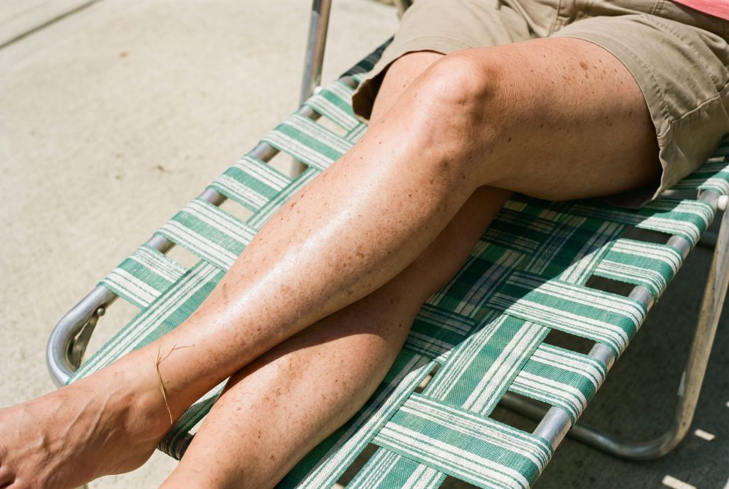
Legs collect pigment for more reasons than most people realize. They get sun in summer and friction year-round, they’re shaved, scratched and bitten, and the lower legs are where circulation problems show up first. That’s why “dark spots on legs” can mean six different things on six different people.
This guide compares the common causes side by side so you can narrow down what you’re looking at, then covers what helps each one and which spots need a professional opinion rather than a cream.
Jump to
Common causes of dark spots on legs
1. Age spots (sun spots)
Age spots, also called liver spots, sun spots or solar lentigines, are flat, round or oval patches that are tan to dark brown and range from freckle-sized to about half an inch (13 millimeters) across (Cleveland Clinic). Ultraviolet light speeds up melanin production, and over years the pigment clusters into spots. They have nothing to do with the liver. They’re more common after 50 and in people with fair skin, a history of bad sunburns or tanning bed use, but anyone can get them. Cleveland Clinic lists the face, hands, neck and arms as typical locations; the common thread is sun exposure. Our explainer on age spots vs. sun spots on legs covers them in detail.
2. Marks left by inflammation (PIH)
Any injury or rash can leave a flat brown, gray or purplish mark behind as it heals. That’s post-inflammatory hyperpigmentation (PIH). On legs, the usual culprits are bug bites, razor nicks and scratches, along with skin conditions such as eczema and psoriasis, which the AAD lists among common triggers (AAD). PIH tends to be more noticeable and longer-lasting on medium and deep skin tones. The good news: once the trigger stops, many of these spots fade on their own, although it can take a while.
3. Seborrheic keratoses
These are the spots that look stuck on. Seborrheic keratoses start as small rough bumps and slowly thicken into waxy, warty growths that can be anything from white to black, though most are brown (AAD). They’re benign and usually painless, though some itch. The key difference from an age spot is texture: you can feel a seborrheic keratosis, while an age spot is completely flat.
4. Diabetic dermopathy (shin spots)
Round or oval, brown or reddish-brown spots on the shins can be a skin sign of diabetes. The AAD describes them as scaly at first, flattening over time and sometimes leaving small dents, with no itch or pain, which makes them easy to mistake for age spots (AAD). The spots themselves are harmless and often fade within 18 to 24 months after diabetes is well controlled. They matter because people who have them face a higher risk of diabetes complications such as nerve damage, and because they sometimes appear in people who don’t yet know they have diabetes.
5. Discoloration from poor circulation
When the veins in the lower legs don’t return blood efficiently (venous insufficiency), the skin can become inflamed and discolored. This is stasis dermatitis. According to the AAD, early signs include itchy, dry, discolored skin, especially over varicose veins, ankle swelling by evening and a heavy or aching feeling in the legs; it typically starts on the inside of the ankles (AAD). Over time, untreated skin can become deeply pigmented and hardened. This isn’t a cosmetic issue, and the AAD stresses that early treatment works best.
6. Capillaritis (pigmented purpura)
Capillaritis causes clusters of tiny red-brown dots, often described as cayenne pepper spots, that can merge into flat patches. They happen when small blood vessels near the surface leak red blood cells, which leave iron-containing pigment behind. The most common form, Schamberg disease, usually appears on the lower legs. DermNet describes it as harmless and mild; it may clear in weeks, come and go, or persist for years (DermNet). Occasionally a medicine or infection is the trigger.
7. Moles and melanoma
Most moles are harmless. But legs deserve particular attention in women: the National Cancer Institute notes that in women, melanoma forms most often on the arms and legs, while in men it’s most often on the trunk, head and neck (NCI). A spot that is changing, or that looks different from the others on your body, needs a dermatologist’s eye (AAD). We explain the warning signs in brown spots on legs: when to get them checked.
The short list
- Flat, tan-to-brown spots on sun-exposed skin are usually age spots.
- Flat marks after bites, nicks or rashes are usually PIH and often fade once the trigger stops.
- Raised, waxy, stuck-on spots are usually seborrheic keratoses.
- Brown shin spots, rust-colored ankle discoloration or cayenne-pepper dots can reflect diabetes, circulation or capillary issues.
- Any spot that changes, bleeds or looks unlike your others should be checked.
Causes compared at a glance
| Cause | What it looks like | Feel | Where on the legs | Next step |
|---|---|---|---|---|
| Age spots | Flat, tan to dark brown, round or oval | Flat | Sun-exposed areas | Sun protection; cosmetic treatment optional |
| PIH | Flat brown, gray or purplish mark where skin was injured | Flat | Wherever bites, nicks or rashes were | Stop the trigger; fading ingredients |
| Seborrheic keratosis | Waxy, warty, white to black | Raised, stuck-on | Anywhere except palms and soles | Usually none; doctor can remove |
| Diabetic dermopathy | Round or oval brown or reddish-brown | Scaly early, then flat or dented | Shins | Ask your doctor about diabetes screening |
| Stasis dermatitis | Discolored, dry, itchy; may turn deeply pigmented | Can harden over time | Inner ankles, lower legs, over varicose veins | See a doctor; treat circulation |
| Capillaritis | Tiny red-brown dots, cayenne-pepper patches | Flat | Lower legs | Usually harmless; get it diagnosed |
| Melanoma | Asymmetric, irregular border, uneven color, changing | Flat or raised | Anywhere; legs are a common site in women | Dermatologist promptly |
Sources: Cleveland Clinic, AAD, AAD, AAD, AAD, DermNet, AAD.
Five questions that narrow it down
You can’t diagnose a spot at home, but you can collect the details a dermatologist will ask about. These five questions sort most leg spots into the right bucket.
- Is it flat or raised? Run a fingertip over it with your eyes closed. Age spots, PIH and capillaritis are flat. Seborrheic keratoses feel waxy or rough and raised.
- Where exactly is it? Shins point toward diabetic dermopathy; inner ankles and skin over varicose veins point toward circulation; the sunniest areas point toward age spots.
- What happened there first? A bite, a nick, a patch of eczema or a scratch that healed into a mark is the classic PIH story.
- Is it one spot or a pattern? Many similar spots that match each other are usually benign. A single spot that looks unlike the rest deserves attention.
- Is it changing? Growth, new colors, bleeding or itching over weeks to months moves a spot straight to the dermatologist list.
Take a clear photo of anything you’re unsure about, with a coin or ruler next to it, and repeat it monthly. That record turns a vague “I think it’s bigger” into something a doctor can use. It also helps with spots you can’t see well, such as the backs of the calves; a hand mirror or a partner helps there too.
What helps fade dark spots on legs
The right approach depends entirely on the cause, which is why diagnosis comes before shopping. For the most common cosmetic causes, age spots and PIH, here’s what actually moves the needle.
Sun protection, every time your legs are out
Sunscreen won’t erase existing spots, but without it, nothing else will work for long, because UV keeps stimulating pigment. Use a broad-spectrum, water-resistant SPF 30 or higher and reapply every two hours outdoors (AAD). Legs are where people skimp: the AAD’s rule of about 1 ounce, a shot glass, covers all exposed skin in a swimsuit, and two legs take a good share of it. For darker skin tones prone to PIH, the AAD suggests a tinted sunscreen with iron oxides, which also block visible light (AAD).
Fading ingredients
Cleveland Clinic lists glycolic acid, kojic acid, vitamin C, niacinamide and salicylic acid as over-the-counter options for hyperpigmentation (Cleveland Clinic), and the AAD adds azelaic acid and retinoids such as retinol and adapalene (AAD). Pick one, use it consistently on the spots or the whole lower leg, and give it at least two to three months. Gentle exfoliating acids also help with the rough, dull texture that often comes with sun-damaged legs; our lactic acid lotion guide explains how.
In-office treatments
For age spots, a dermatologist may suggest prescription creams, chemical peels, cryotherapy (freezing), dermabrasion or laser treatment (Cleveland Clinic). These work faster than creams but carry their own risk of temporary or lasting pigment changes, particularly on deeper skin tones, so experience matters when choosing who treats you.
Treating the cause
For shin spots linked to diabetes, getting blood sugar under control is what helps them fade (AAD). For stasis dermatitis, the fix is improving circulation, which is a job for your doctor rather than a fade cream (AAD). For capillaritis, DermNet notes that compression stockings can help lower-leg cases and that a doctor may review medicines as possible triggers (DermNet).
How long it takes
Expect months, not weeks. The AAD says a spot a few shades darker than your natural skin color usually fades in 6 to 12 months once its cause is gone, and deeper discoloration can take years (AAD). Cleveland Clinic gives a similar range for treatment: a few months to a year (Cleveland Clinic).
When to see a dermatologist
Get it looked at if
- a spot shows any of the ABCDE signs: asymmetry, an irregular border, several colors, a diameter over about 6 mm, or it’s evolving
- a spot looks different from your other spots (the “ugly duckling”)
- a spot bleeds, itches persistently or appeared suddenly
- you have brown spots on your shins and haven’t been checked for diabetes
- your ankles swell, your legs feel heavy, or the skin near varicose veins is discolored, itchy or hardening
- you’d like age spots treated and want to know which option suits your skin tone
ABCDE criteria from the AAD. Early detection matters: the AAD reports that the average five-year survival rate for melanoma caught before it spreads to the lymph nodes is 99% (AAD). The AAD also notes that in people with darker skin tones, melanoma is often diagnosed later, when it’s harder to treat.
Frequently asked questions
Why am I getting dark spots on my legs?
The most common reasons are sun exposure (age spots) and marks left by bites, shaving nicks or rashes (PIH) (Cleveland Clinic; AAD). Spots concentrated on the shins or around the ankles can point to diabetes or circulation problems and are worth showing a doctor.
Are dark spots on legs a sign of diabetes?
Some can be. Round, brown or reddish-brown spots on the shins, called diabetic dermopathy, are a recognized skin sign of diabetes. They’re harmless themselves but can signal a higher risk of complications (AAD).
Can dark spots on legs be cancer?
Rarely, but it happens, and legs are a common melanoma site in women (NCI). Use the ABCDE signs and see a dermatologist for any spot that is new and unusual or changing (AAD).
How can I get rid of dark spots on my legs?
For age spots and PIH: daily sunscreen plus a fading ingredient such as azelaic acid, glycolic acid, niacinamide, vitamin C or a retinoid, used consistently for months (AAD; Cleveland Clinic). A dermatologist can offer peels, freezing or lasers for faster results.
Do dark spots on legs go away on their own?
Marks from bites or rashes often fade on their own within 6 to 12 months once the cause stops (AAD). Age spots generally don’t fade without treatment, and capillaritis can come and go (DermNet).
About the author
Rachel Moreno writes Body Skin Insider's guides to crepey skin, discoloration, dry skin and the ingredients that help, with a focus on the skin below the chin. Each guide lists its sources and spells out when a spot or change should be seen by a dermatologist.
How we put this together
We built this guide from 11 sources, including Cleveland Clinic and the American Academy of Dermatology. Key claims link to the source that supports them.
Sources
- Cleveland Clinic. “Liver Spots: Causes, Symptoms and Treatment.” Cleveland Clinic. my.clevelandclinic.org
- Cleveland Clinic. “Hyperpigmentation: What it is, Causes and Treatment.” Cleveland Clinic. my.clevelandclinic.org
- American Academy of Dermatology. “How to fade dark spots in darker skin tones.” AAD. aad.org
- American Academy of Dermatology. “Diabetes: 12 warning signs that appear on your skin.” AAD. aad.org
- American Academy of Dermatology. “Stasis dermatitis: Signs and symptoms.” AAD. aad.org
- DermNet. “Capillaritis (pigmented purpura).” DermNet. dermnetnz.org
- American Academy of Dermatology. “Seborrheic keratoses: Signs and symptoms.” AAD. aad.org
- American Academy of Dermatology. “Melanoma: Signs and symptoms.” AAD. aad.org
- National Cancer Institute. “Melanoma Treatment (PDQ): Patient Version.” NCI. cancer.gov
- American Academy of Dermatology. “Melanoma” (skin cancer statistics). AAD Newsroom. aad.org
- American Academy of Dermatology. “How to apply sunscreen.” AAD. aad.org
General information, not medical advice. For a spot that’s new, changing or bleeding, see a dermatologist.